

Traumatic Vertebral Fracture
Emergency stabilization of a T12 fracture — precise, atypical screw trajectories navigated with the C-arm and staff already in the room.
Patient Overview
Age
60 years old
Sex
Male
Diagnosis
T12 fracture
Surgery Details
Procedure
T11-L1 stabilization to restore neurologic function and return to daily activity
Levels with fixation
T11-L1
Fixation type
Pedicle screw fixation
Patient Background and Diagnosis
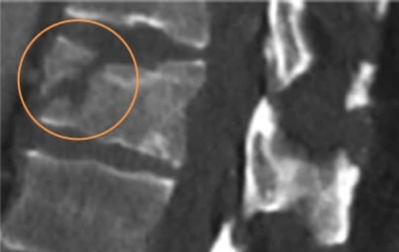
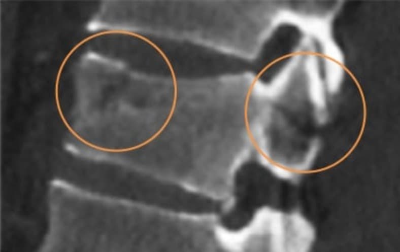
This patient was a 60-year-old male working on an oil rig when a 400-pound cylindrical pipe fell and struck him on the back. He was transported to the hospital by helicopter. On assessment, he presented with a T12 fracture and an associated neurological deficit — the fracture involved the vertebral body, facets, and pars interarticularis.
Surgical Treatment
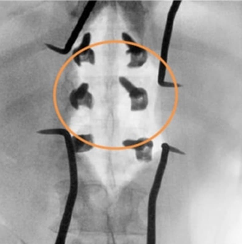
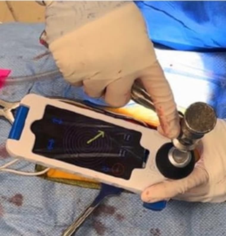
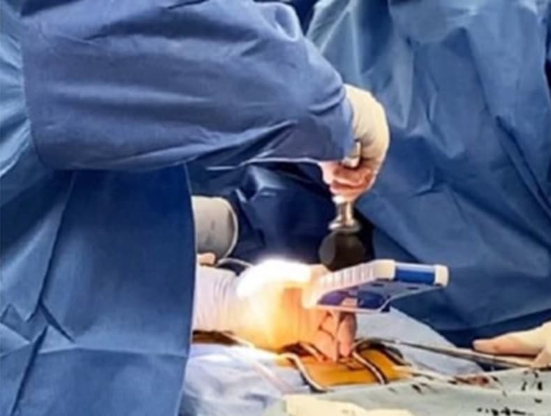
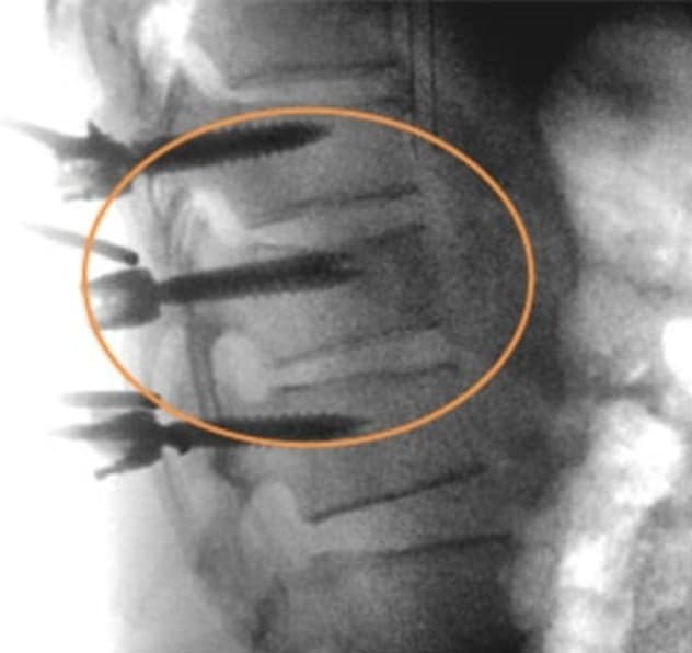
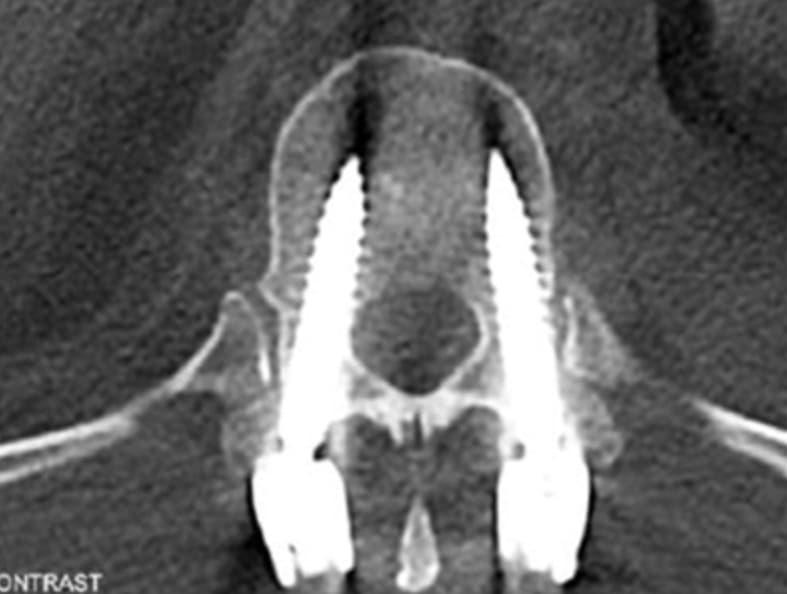
This was a T11-L1 stabilization to restore neurologic function and return the patient to daily activity. The screws at T12 had to follow diverging medial and sagittal trajectories to avoid the fractured elements — the surgeon planned and placed each one under Bolt guidance.
Surgical Experience
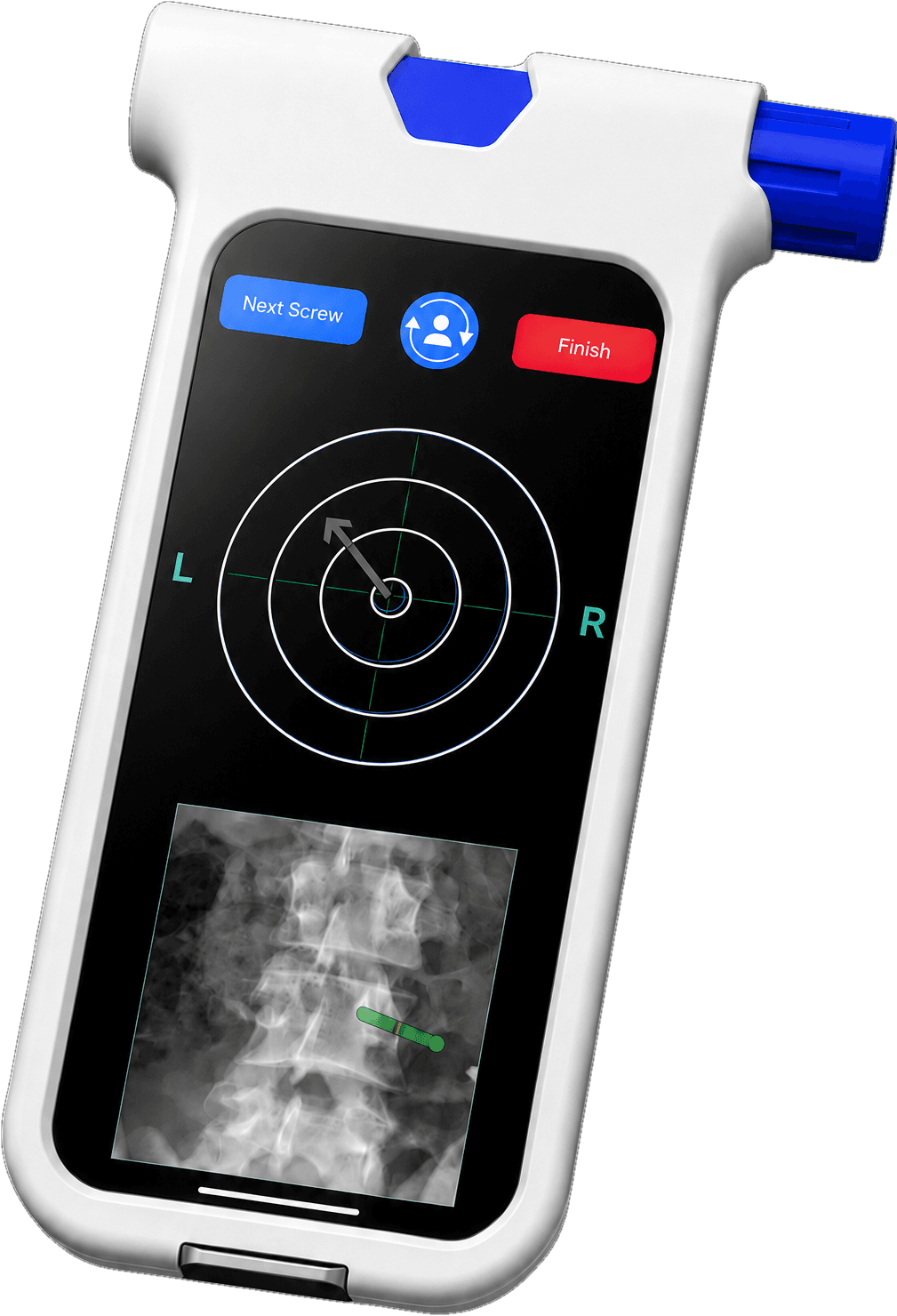
In an emergency setting, the surgeon planned and placed each screw precisely using a standard C-arm and the patient's diagnostic imaging — no O-arm, no reference frame, no outside technician. Bolt navigated the atypical trajectory with the staff and equipment already in the room: a practical, low-radiation alternative to legacy navigation for a challenging case.
Outcome
The patient was discharged four days after surgery with full neurologic function, and returned to full activity in under three months.
SeeBoltinyourOR.
TalkwithaclinicalspecialistabouthowBoltfitsthecasesyourun—physician-controlled,withnocapitalexpense.