

Decompression and Fusion
An L4-5 laminectomy and posterior fusion — four pedicle screws planned and placed level by level under Bolt guidance.
Patient Overview
Age
69 years old
Sex
Male
Surgery Details
Procedure
L4-5 laminectomy and posterior fusion
Levels with fixation
L4-5
Fixation type
Pedicle screws
Operative Workflow
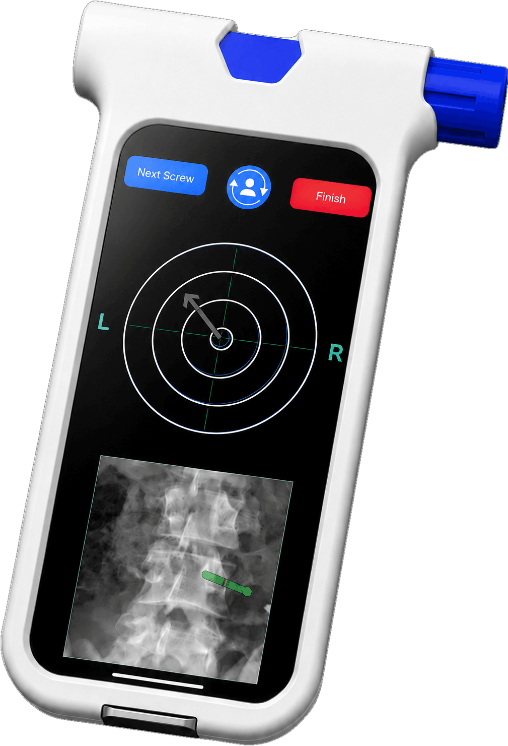
The patient was positioned prone on a standard OR table and prepped and draped in standard sterile fashion. The lumbar MRI, taken two months before surgery, was opened on the OR computer. The screen was leveled with the Bolt Navigation Unit (BNU), and axial images of L4 and L5 were acquired. A C-arm (OEC 9900, GE, Chicago, IL) was draped and positioned, and a lateral lumbar spine X-ray was taken. The L4 and L5 images from the C-arm were acquired separately with the BNU, which was then placed in a sterile drape and passed to the scrub technician.
A 7 cm incision exposed the L4 and L5 spinous processes and laminae, and a decompressive laminectomy was performed at both levels. Using a lateral X-ray to locate the entry point of the right L4 pedicle, the surgeon planned the screw trajectory on the BNU. With the trajectory set, the BNU was attached to the gearshift probe using the single-use case, and — under Bolt guidance — the probe was advanced through the pedicle along the planned path into the vertebral body. The path was tapped (Alphatec Spine, Inc., Carlsbad, CA) and a pedicle screw placed. This was repeated for the right L5, left L4, and left L5 pedicles. The posterior lateral gutters were prepped, the screws connected with rods, and the procedure completed.
Perioperative
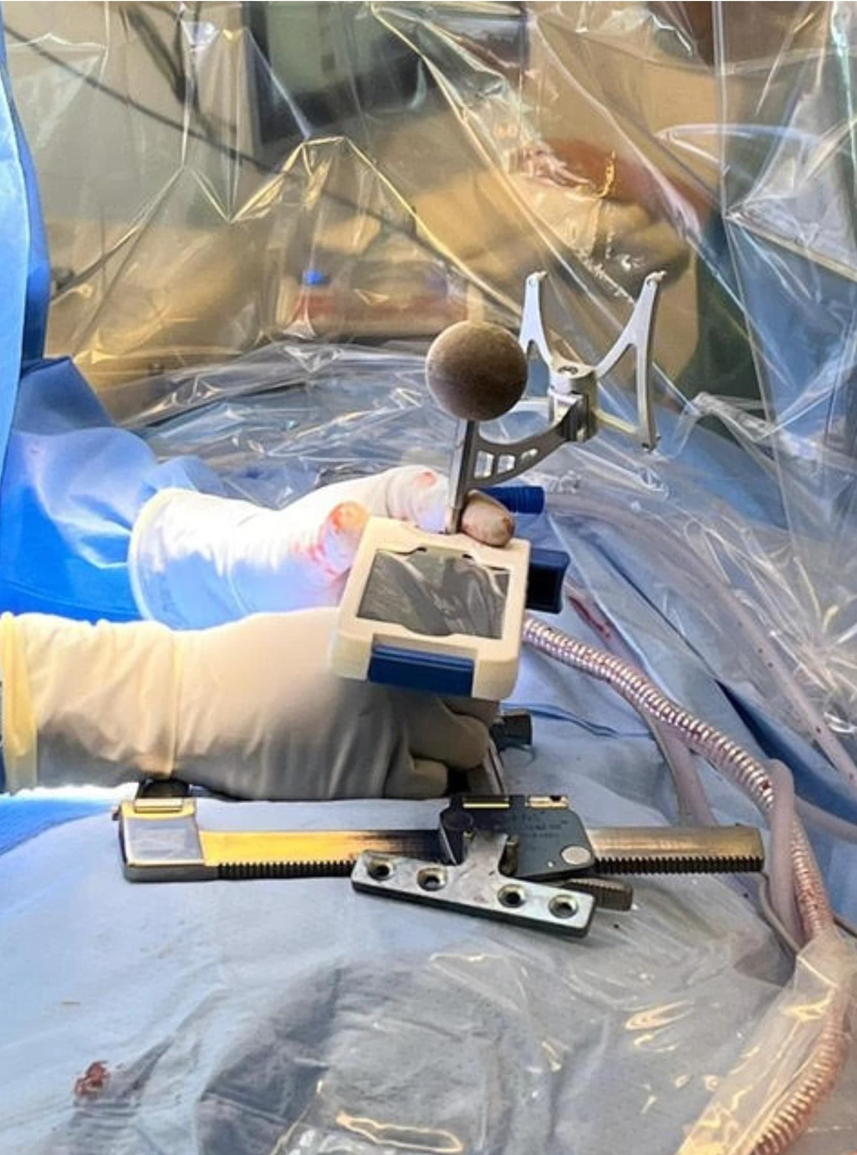
Post-op
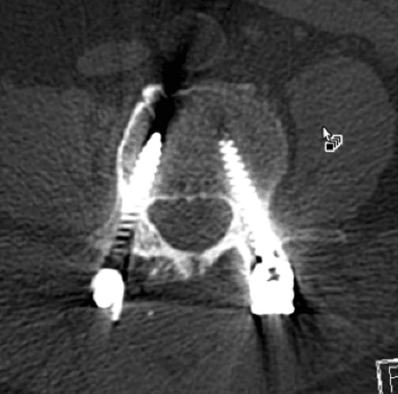
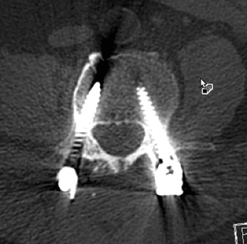
Post-op axial CT: all four L4-5 pedicle screws accurately placed (two views).
Outcome Summary
All four pedicle screws placed accurately under Bolt guidance.
SeeBoltinyourOR.
TalkwithaclinicalspecialistabouthowBoltfitsthecasesyourun—physician-controlled,withnocapitalexpense.