Cortical Bone Trajectory
Precise cortical bone trajectory with minimal radiation — four CBT screws placed under Bolt® guidance from a single lateral and single A/P X-ray.
Patient Overview
Age
60 years old
Sex
Male
Diagnosis
L3-4 stenosis, mild spondylolisthesis and instability
Surgical Treatment
The patient underwent an L3-4 decompression and interbody fusion to relieve pain, stabilize the segment, and restore quality of life. Dr. Thompkins elected a posterior Cortical Bone Trajectory (CBT) technique: optimal screw purchase into high-density bone, a smaller incision, less muscle retraction, lower blood loss, shorter surgical time, and directional avoidance of neural anatomy.1 In CBT, a precise trajectory is critical — to achieve optimal cortical purchase and prevent a breach of the lateral mass of the vertebral body.
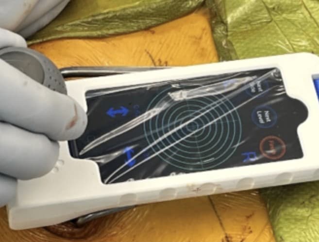
Dr. Thompkins decompressed the compromised anatomy, placed four CBT screws under Bolt guidance, and implanted two TLIF cages at L3-4: a porous titanium TLIF cage and a structural curved allograft.
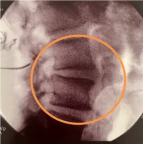
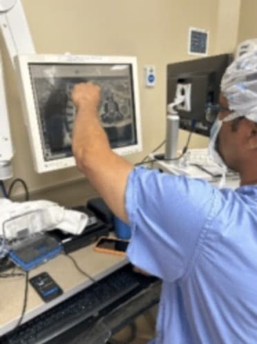
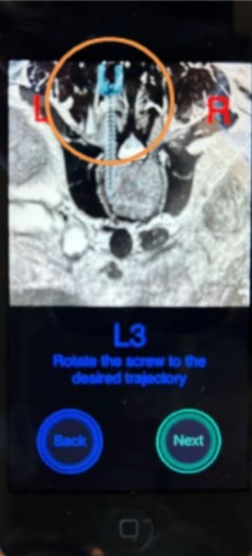
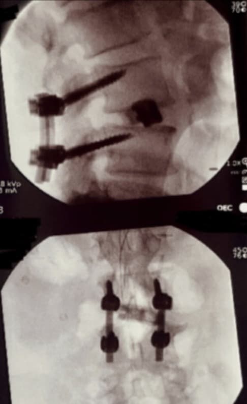
His technique for cortical trajectory with Bolt maximizes accuracy and minimizes radiation. Caspar pins are placed at the selected entry point for each screw; with the pins in place, a single lateral X-ray and a single A/P X-ray are acquired. Dr. Thompkins uses the pins as reference points to assess each screw's entry point, angulation, and congruency, then plans any trajectory modifications directly in Bolt. In this case, he corrected the angle of the top right pin during planning and — as the post-placement X-ray confirms — achieved optimal screw placement.
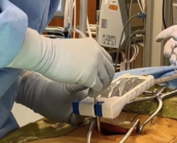
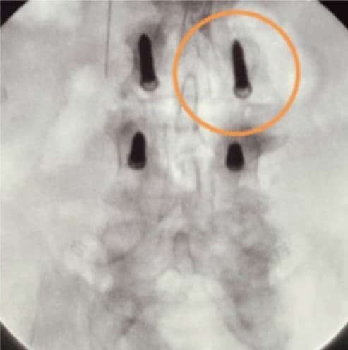
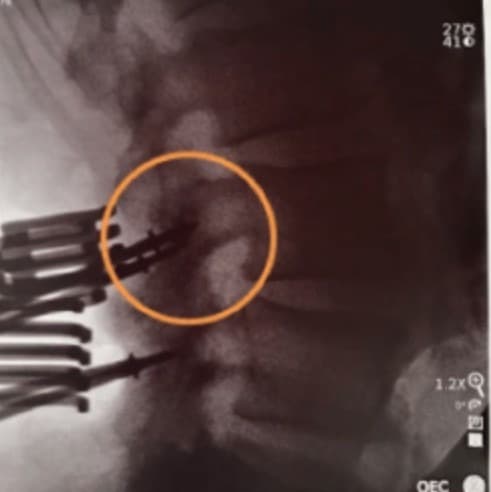
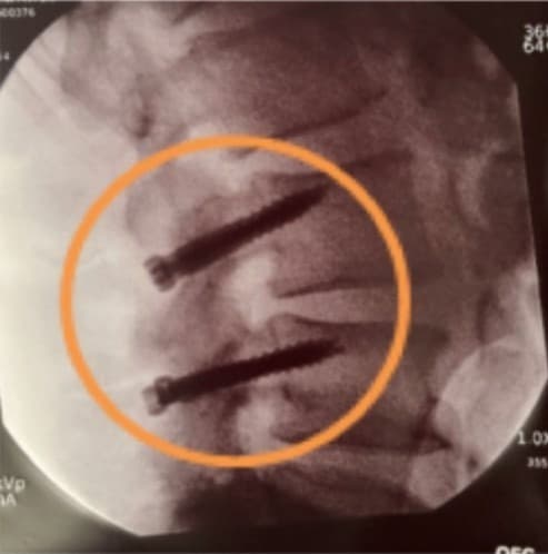
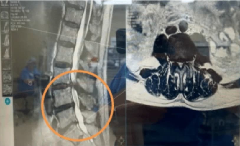
CBT demands increased accuracy because of the narrow placement corridor. Here, Bolt enabled precise placement of cortical screws through a confined midline access, with a straight-on, slightly cranial, divergent angulation away from the neural elements — fixation through the medial and lateral cortex for advanced bony purchase. Bolt let Dr. Thompkins achieve the benefits of cortical trajectory quickly, with minimal radiation exposure.2,3The system leverages limited intraoperative X-rays and the patient's existing pre-operative MRI or CT to deliver efficient, precise, low-radiation navigation for this minimally invasive approach.
Outcome
The patient was discharged 3 days after surgery. He returned to full activity less than 3 months after the procedure.
- 1. Gutierrez J, Erwood A, Malcom J, Grogan D, Greven A, Gary M, Rodts G, Stricsek G, Refai D. "Comparing Cortical Bone Trajectory and Traditional Pedicle Screws in Transforaminal Lumbar Interbody Fusion: A Retrospective Cohort Study of One-Year Outcomes." Cureus. 2023 Aug;15(8).
- 2. Zhang J, Weir V, Fajardo L, Lin J, Hsiung H, Ritenour ER. "Dosimetric characterization of a cone-beam O-arm imaging system." J Xray Sci Technol. 2009;17(4):305-317.
- 3. Mendelsohn D, Strelzow J, Dea N, et al. "Patient and surgeon radiation exposure during spinal instrumentation using intraoperative computed tomography-based navigation." Spine J. 2016;16(3):343-354.
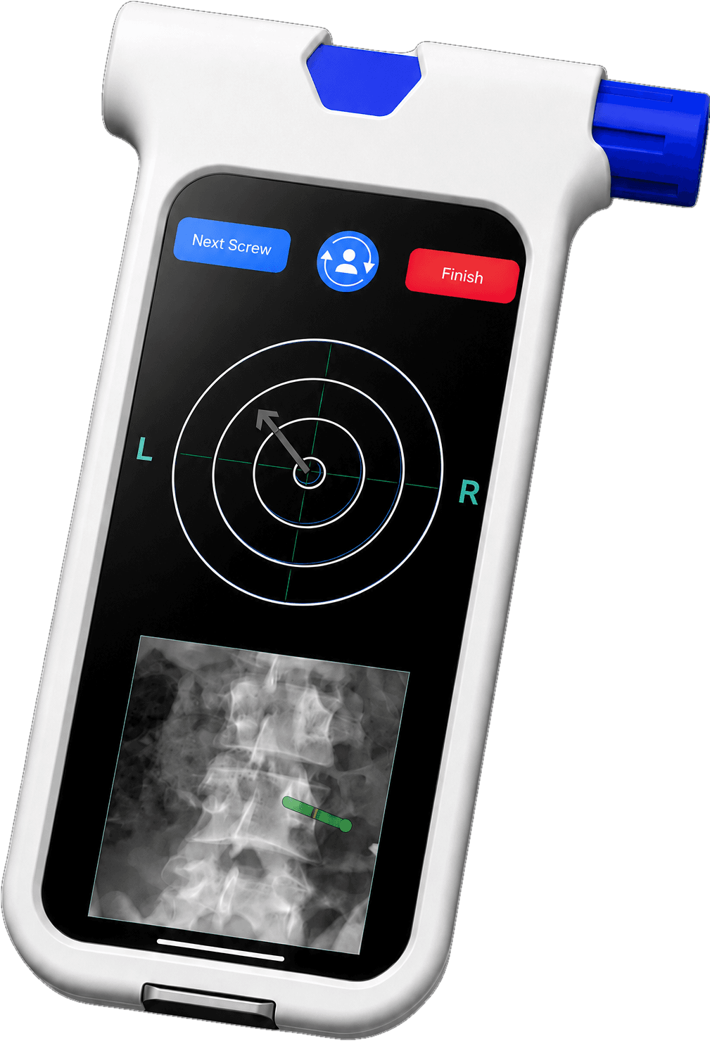
SeeBoltinyourOR.
TalkwithaclinicalspecialistabouthowBoltfitsthecasesyourun—physician-controlled,withnocapitalexpense.